High-resolution X-ray tomography reveals how butterflies form their iridescent wing scales
It has only been one year since the material scientists around Prof. Erdmann Spiecker from the Center for Nanoanalysis and Electron Microscopy (CENEM) at the FAU Erlangen-Nürnberg had been granted funding for one of the World’s best X-ray microscopes, and they could already contribute with fascinating 3D analyses to unravel an open question in butterfly research. Their results have recently been published in the renowned scientific journal Science Advances.
Fascinating colors generated by photonic crystals
Who is not fascinated by the wonderful iridescent colors of butterfly wings? If you try to understand this phenomenon, you will find out that most often the color is not generated by pigments but rather by periodic structures made of chitin, a structure-forming polysaccharide. These so-called photonic crystals give rise to structural color by only reflecting specific wavelengths of the incoming solar spectrum. The resulting color is not random but rather serves as camouflage or signaling. But how do millions of these photonic crystals form within the tiny scales of butterfly wings? The opinions of scientists differ in this matter.
“Time-frozen” stages of crystal growth
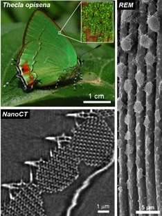
In a cooperation with two leading experts in butterfly research, Dr. Bodo Wilts from University of Fribourg/Switzerland and Dr. Gerd Schröder-Turk from Murdoch University in Perth/Australia, as well as with Dr. Stephen Kelly from Zeiss-Xradia the material scientists from Erlangen employed different high-resolution microscopy techniques to reveal the formation mechanism of these photonic crystal structures. The green butterfly Thecla opisena, which has its habitat in the Neotropics from Mexico to Venezuela, features separated photonic crystal domains which increase in size from the base to the tip of the wing scales. This is a so-far unique characteristic, distinct to other butterflies. The scientists interpret this finding in such a way that the photonic crystal growth got “time-frozen” at different stages of the metamorphosis. Hence, detailed microscopic analyses can be employed to obtain important insights into the formation processes of these crystals.
X-ray tomography resolves finest details
“High-resolution X-ray tomography could provide essential findings for a deeper understanding of the formation mechanisms”, explained Prof. Erdmann Spiecker. The scientists assume that nascent chitin is extruded into a ‘casting mould’ made of membranes. “The unique possibility of X-ray tomography to analyze the 3D structure of entire wing scales was used to clarify from where the chitin was finally extruded.” Dr. Benjamin Apeleo Zubiri, who analyzed the 3D data in detail, is amazed: “The resolution of the reconstructed tomograms is so high, that not only this question could be clarified, but also the chirality (handedness) of each single photonic crystal could be identified.” So far, this was only possible by using electron tomography, as the researchers from CENEM could demonstrate in an earlier study. However, for the electron tomography investigation, the wing scales needed to be cut into little segments, which caused serious disadvantages.
Modern materials science using X-ray tomography
Even though the material scientists have already been investigating these butterfly wing scales for several years, this is a rather ‘exotic‘ application at CENEM. “We typically employ advanced microscopy and tomography techniques to enhance our knowledge about modern functional and energy materials and to optimize their properties for applications”, explained Prof. Erdmann Spiecker. “The new X-ray microscope will also enable inimitable insights in these areas, for example regarding the investigation of porous structures for catalytic applications or the search for tiny failures in turbine materials.” But, also photonic crystals are closely related to modern materials science. These intriguing 3D structures with their unique optical properties may serve as prototypes for novel functional materials with application in, e.g., photovoltaics.
This press release was originally published in German language here.
Further information:
Prof Dr Erdmann Spiecker
Tel.: 09131/85-28603
erdmann.spiecker@fau.de
em.tf.fau.de, cenem.fau.de
Publication:
“Butterfly gyroid nanostructures as a time-frozen glimpse of intracellular membrane development”, Bodo D. Wilts, Benjamin Apeleo Zubiri, Michael A. Klatt, Benjamin Butz, Michael G. Fischer, Stephen T. Kelly, Erdmann Spiecker, Ullrich Steiner and Gerd E. Schröder-Turk, Science Advances 3, e1603119 (2017).