Nanoparticulate structures and nanoporous materials
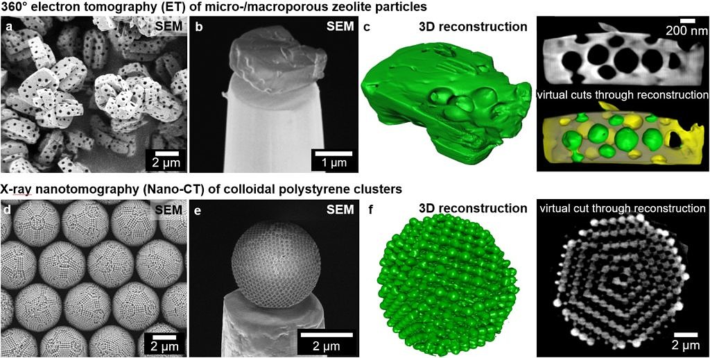 Two examples of scale-bridging and complementary tomography techniques on nanoparticulate structures and nanoporous materials: (upper row) 360° electron tomography (ET) of micro-/macroporous MFI-type zeolite particles and (bottom row) X-ray nanotomography (Nano-CT) of magic colloidal clusters composed of >1000 polystyrene (PS) balls: (a, d) SEM overview images of several particles/clusters; (b, e) single particle/cluster on tomography tip plateaus; (c, f) Renderings of and further virtual cuts through 3D reconstructions.
Quantitative knowledge of the 3D structure of particles and particle aggregates down to nanometer scale is essential for a fundamental understanding, modelling and optimisation of optical properties like, e.g., plasmon resonances and structural colour. Similarly, quantitative knowledge of the 3D pore structure of particles, thin films and stationary phase materials is crucial for understanding, modelling and optimising for instance nanoparticle separation by chromatographic processes or gas transport properties for catalytic applications. Electron microscopy and X-ray microscopy techniques play a key role for structural characterisation of nanoparticles, particle ensembles and nanoporous materials and are extensively used for evaluation of synthesis routes and particle and pore design principles.
At IMN, we perform cutting-edge electron microscopy, electron tomography and Nano-CT characterisation of nanoparticulate and nanoporous materials by using the state-of-the-art equipment of the Center for Nanoanalysis and Electron Microscopy (CENEM).
The research in this area is partly funded by the follow-up institution at FAU of the DFG Cluster of Excellence EXC 315 “Engineering of Advanced Materials”. Moreover, this research area is funded by the DFG within SFB1141 “Design of Particulate Products” as well as SFB1452 “Catalysis at liquid interfaces”. Within these projects, we have built up a remarkable track record throughout the last years in the field of nano-tomography including Nano-CT, electron tomography and FIB/SEM tomography, since these 3D imaging techniques are often indispensable to unravel important properties of the studied class of material.
Current members
Current PhD students


Allison Götz, M.Sc.
91058 Erlangen
- Phone number: +49 9131 85-70380
- Email: alexander.goetz@fau.de
- Website: http://www.em.techfak.uni-erlangen.de

Graduated PhDs
Dr.-Ing. Stefanie Spallek
Dr.-Ing. Benjamin Apeleo Zubiri
Dr.-Ing. Thomas Przybilla
Dr.-Ing. Janis Wirth
- , , , , , , , :
Scale-Bridging 3D Analysis of Micro-/Macroporous Zeolite Particles Using X-Ray Nano-Tomography and Electron Tomography
DOI: 10.1017/S143192761900271X
URL: https://www.cambridge.org/core/journals/microscopy-and-microanalysis/article/scalebridging-3d-analysis-of-micromacroporous-zeolite-particles-using-xray-nanotomography-and-electron-tomography/08B01EC1985E6D09CCA6940D31E3B246
BibTeX: Download - , , , , , , :
Scale-Bridging 3D-Analysis of Colloidal Clusters Using 360° Electron Tomography and X-Ray Nano-CT
DOI: 10.1017/S1431927619002691
URL: https://www.cambridge.org/core/journals/microscopy-and-microanalysis/article/scalebridging-3danalysis-of-colloidal-clusters-using-360-electron-tomography-and-xray-nanoct/0FD7C5C09F258792F594A66FAC2E1A7B
BibTeX: Download - , , , , , , :
Free Energy Landscape of Colloidal Clusters in Spherical Confinement
In: Acs Nano (2019)
ISSN: 1936-0851
DOI: 10.1021/acsnano.9b03039
BibTeX: Download - , , , , , , :
Structural Analysis of Liquid Metal Catalysts in Porous Silica Utilizing Nano-CT and Analytical Transmission Electron Microscopy
DOI: 10.1017/S1431927619002848
URL: https://www.cambridge.org/core/journals/microscopy-and-microanalysis/article/structural-analysis-of-liquid-metal-catalysts-in-porous-silica-utilizing-nanoct-and-analytical-transmission-electron-microscopy/B715B14D19AFC5BDE694C8CDEF030CD5
BibTeX: Download - , , , , , , :
Magic number colloidal clusters as minimum free energy structures
In: Nature Communications 9 (2018), Article No.: 5259
ISSN: 2041-1723
DOI: 10.1038/s41467-018-07600-4
BibTeX: Download - , , , , , :
Correlative 3D-Characterization of Liquid Metal Catalysts (LMC) utilizing X-ray and Analytical Electron Microscopy
14th International Conference on X-ray Microscopy (XRM2018) (Saskatoon, Saskatchewan, Canada, August 19, 2018 - August 24, 2018)
In: Microscopy Society of America 2018 (ed.): Microscopy & Microanalysis 2018 2018
DOI: 10.1017/S1431927618014976
BibTeX: Download - , , , , , :
Coexistence of both gyroid chiralities in individual butterfly wing scales of Callophrys rubi
In: Proceedings of the National Academy of Sciences of the United States of America 112 (2015), p. 12911-12916
ISSN: 0027-8424
DOI: 10.1073/pnas.1511354112
BibTeX: Download - , , , , , , , , , :
Transfer of Individual Micro- and Nanoparticles for High-Precision 3D Analysis Using 360° Electron Tomography
In: Small Methods 2 (2017), p. 1700276
ISSN: 2366-9608
DOI: 10.1002/smtd.201700276
URL: http://onlinelibrary.wiley.com/doi/10.1002/smtd.201700276/abstract
BibTeX: Download - , , , , , , , , , , :
Three-dimensional and quantitative reconstruction of non-accessible internal porosity in hematite nanoreactors using 360 degrees electron tomography
In: Microporous and Mesoporous Materials 246 (2017), p. 207-214
ISSN: 1387-1811
DOI: 10.1016/j.micromeso.2017.03.028
BibTeX: Download