
Self-assembly processes play an important role in nature and often result in complex structures, e.g. in chlorophyll or in living organisms. Compact clusters of elemental particles can be shown to be of practical relevance, and are found in atomic nuclei, nanoparticles or viruses.
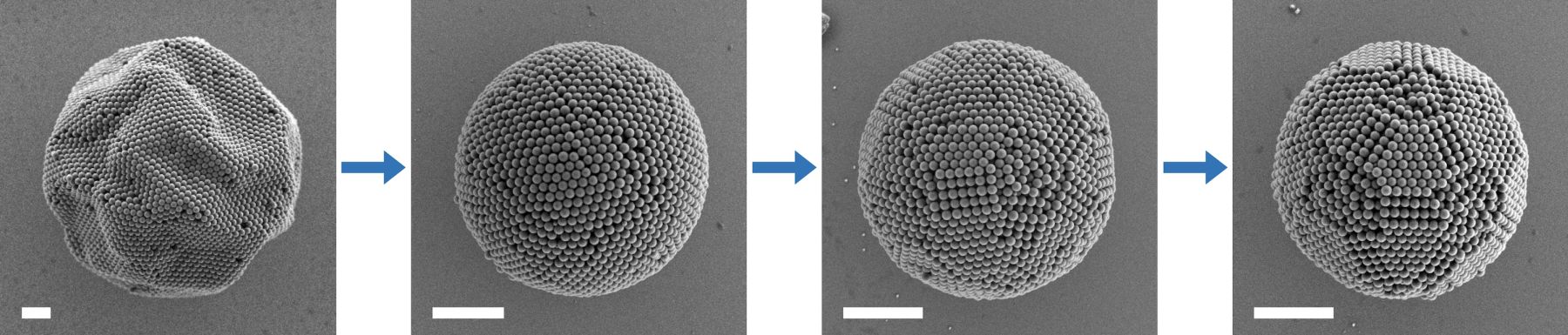 Self-assembly of magic number colloidal clusters takes place in several steps. It is striking to see how several thousand individual particles find optimal positions in a well-defined structure. (scale bar: 2 micrometres) (Image: FAU/Junwei Wang)
An interdisciplinary team of researchers at the FAU led by Prof. Dr. Nicolas Vogel (Institute of Particle Technology), and Prof. Dr. Michael Engel (Institute for Multiscale Simulation) – both from the Department of Chemical and Biological Engineering – together with the materials science expert Prof. Dr. Erdmann Spiecker (Institute of Micro- and Nanostructure Research, IMN) have decoded the structure and the process behind the formation of one class of such highly ordered clusters. Vogel was responsible for synthesis, Spiecker for structure analysis and Engel for modelling clusters from colloidal polymer balls.
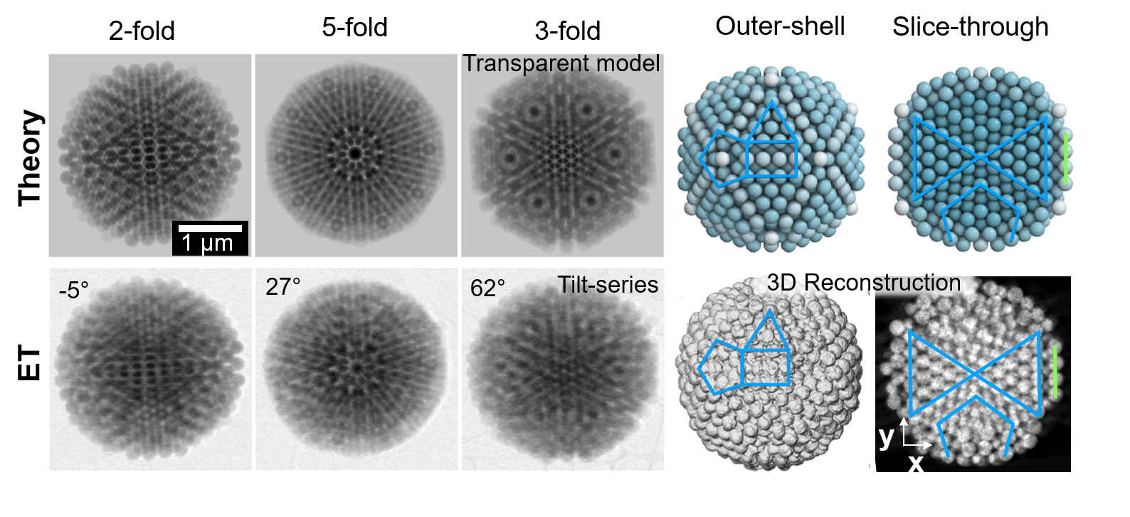 Electron tomography confirmation of model: (bottom row – left) Bright-field (BF) scanning transmission electron microscopy (STEM) images from a tomographic tilt series under specific tilt angles of a magic number colloidal cluster viewing along two-fold, five-fold, and three-fold axes, characteristic for icosahedral symmetry. (bottom row – right) Electron tomography (ET) reconstruction provides a three-dimensional visualization of characteristic surface features of triangles with four particles at side, rectangles of three and four particles at width and length, and pentagon with three particles at side (indicated in blue). Cross-sectional views through the reconstruction at the central plane reveal the characteristic internal structure. A section of the Mackay core is marked by two blue triangles. The vertex region is marked by its characteristic pentagon. The second anti-Mackay shell is indicated by a green line segment. (top row) The experimental images show excellent agreement with model reconstructions using semitransparent spheres mimicking the STEM imaging process, external and cross-sectional views. (Image: FAU/Junwei Wang)
Using high resolution microscopy to show the surface of such clusters does not provide sufficient proof of these symmetries. Even if the surface of a cluster appears highly ordered, that is no guarantee that the particles inside the cluster are arranged as expected. To verify this, PhD student Thomas Przybilla and Dr. Benjamin Apeleo Zubiri from the research team of Prof. Dr. Erdmann Spiecker used electron tomography to elucidate the precise 3D arrangement of these ordered clusters. The researchers from IMN can build on almost 10 years of experience in the development and application of electron tomography techniques. Using the advanced capabilities of the double-corrected Titan transmission electron microscope installed at the Centre for Nanoanalysis and Electron Microscopy (CENEM) the team already employed electron tomography for 3D structure analysis of quantum dots, nanoporous metals foams, mesoporous particles and natural photonic crystals to name only a few applications.
The findings of the interdisciplinary collaboration on magic colloidal clusters have been published in the current issue of the journal Nature Communications
Further information on how magic clusters are formed can be found in the full FAU press release (here).
All images subject to CC BY 4.0 licence