Correlative Nano-CT and FIB Tomography: the paper published in Advanced Engineering Materials is now online!
A paper by Malte Lenz, Janis Wirth, and colleagues, entitled “Correlative Nano‐Computed Tomography and Focused Ion‐Beam Sectioning: A Case Study on a Co‐Base Superalloy Oxide Scale” was recently published in Advanced Engineering Materials.
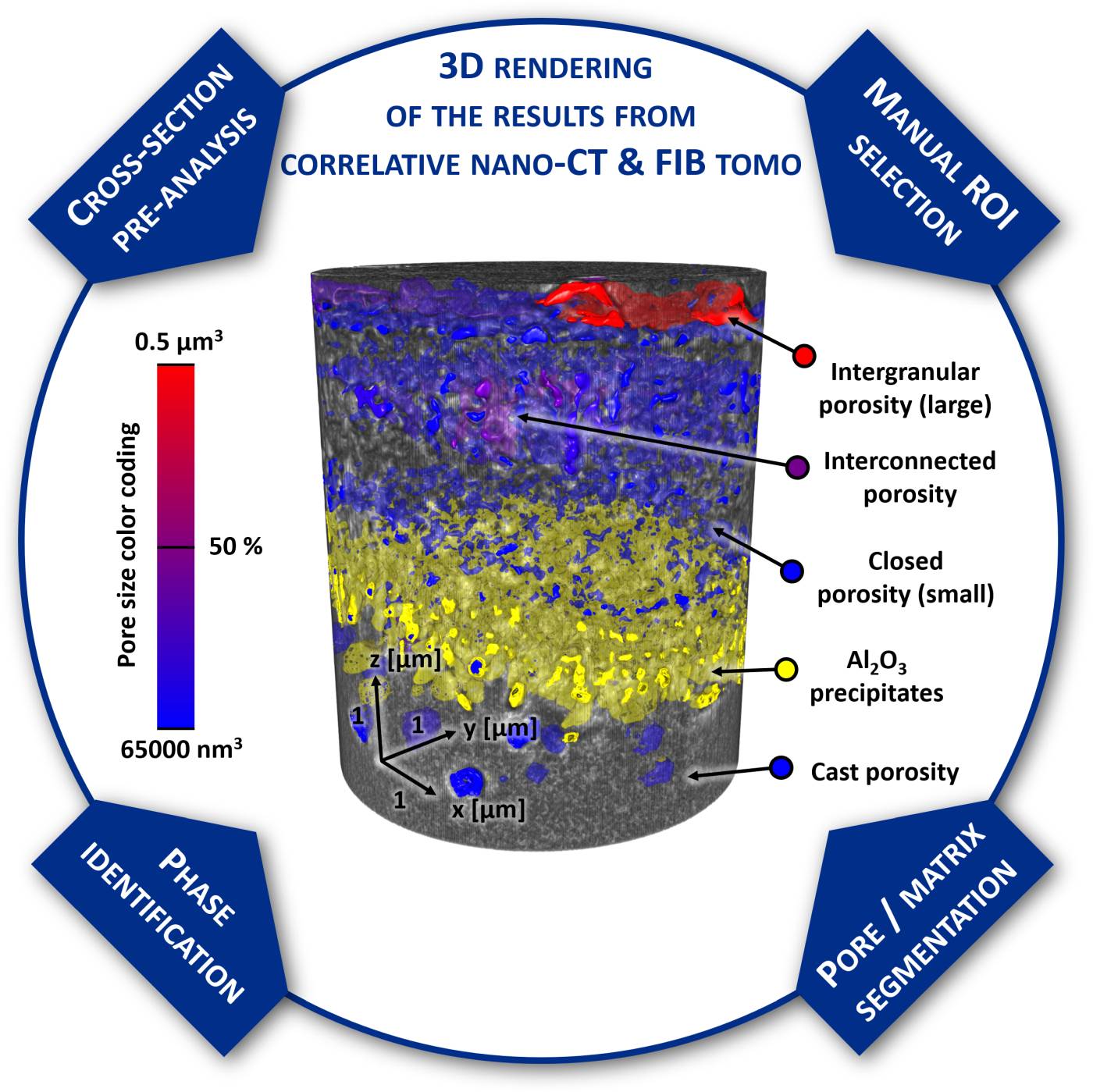
In this work, a correlative tomography approach combining laboratory nano X-ray computed tomography, focused ion beam tomography and energy-dispersive X-ray spectroscopy is applied for the characterization of multi-layered oxide scales of Co-base superalloys regarding their 3D morphology and chemical composition. One of the grand challenges in nano-tomography is how to confidently and properly interpret the contrast (i.e., gray levels) of the 3D tomogram datasets in terms of the physical objects in the sample (e.g., voids, precipitates of different phases). Theoretically, one can simulate the image/contrast formation for simple objects. This is however very difficult to validate for complex scenarios like in the current case of superalloy oxide scales comprising multinary phases. The “tomo team” at WW9/IMN has contributed in the last years to the development of an experimental approach: correlating the multi-dimensional information from different tomographic techniques from the identical sample, i.e., correlative microscopy. The combination of complementary three-dimensional imaging techniques allows for a precise and reliable segmentation of the pore space, oxide precipitates and different phases of the initial material. Such information is fundamental to a microscopic understanding of oxidation in this new class of superalloys and key to improvement of oxidation resistance in the next-generation high-temperature materials.
It’s worth to highlight that two of the contributing authors, Jan Rosiwal and Nadine Buchinger, were student research assistants in the nano-tomography and HT-materials working groups of the IMN and also performed their Bachelor theses on this topic. Their contributions to this work has been already recognized by receiving best poster awards (see news articles here and here) during participating in international conferences.
Congratulations!