Scientists Unveil a New Window into Nanoscale Worlds: Revealing the 3D Atomic Structures of Complex Materials
In a milestone study, IMN scientists have demonstrated a novel technique that uncovers the three-dimensional structures of nanomaterials, including those made of light elements that have eluded detection until now. This advance, which utilizes ptychographic electron tomography, showcases a new tool to aid our understanding of the nanoscale realm, with wide-ranging implications for materials science and structural biology.
Understanding the atomic structure of materials is crucial for deciphering their properties and functions. Traditional methods like X-ray crystallography and conventional electron microscopy have provided insights into crystalline structures and biomolecules. However, these techniques often fall short when it comes to heterogeneous nanomaterials or those containing light elements, which are sensitive to the high-energy beams used for imaging.
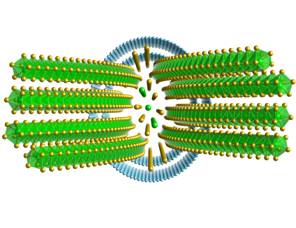
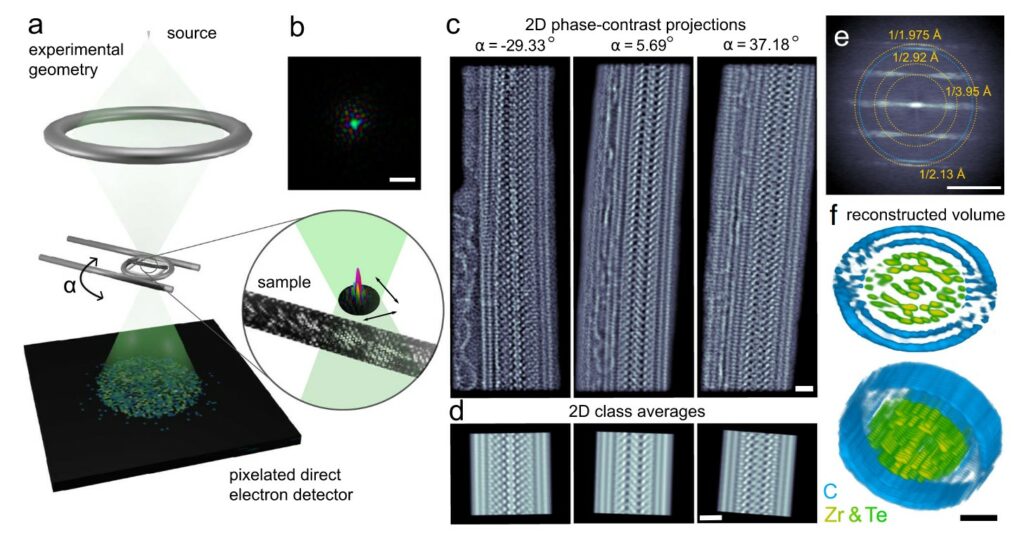
Enter ptychographic electron tomography, a method that has successfully mapped the atomic structure of a double wall-carbon nanotube encapsulating a complex ZrTe (zirconium telluride) structure. This technique relies on collecting millions of diffraction patterns from different angles, which are then synthesized into a three-dimensional volume at atomic resolution. Remarkably, it revealed a Zr11Te50 structure with a previously unobserved ZrTe2 phase in the core, showcasing its ability to illuminate the intricate details of nanomaterials.
Ptychographic atomic electron tomography (PAET) of a complex nanostructure.
This opens up new avenues for the exploration of materials that were previously beyond our reach. Understanding these structures is essential for designing novel materials with desired properties, such as increased strength, improved conductivity, or specific chemical reactivity.
The practical applications of this technology are vast. In materials science, it enables the development of new nanomaterials with tailored properties for use in electronics, energy storage, and catalysis. In structural biology, it could lead to a deeper understanding of the molecular machinery of life, aiding in the development of new drugs and therapies.
Moreover, this method represents a leap forward in terms of dose efficiency and accuracy. Previous approaches required averaging the structures of thousands of identical particles or could not adequately visualize light elements. Ptychographic electron tomography overcomes these limitations, providing a clear, detailed view of the sample at the atomic level.
This breakthrough was made possible through the collaboration of scientists from various disciplines, underscoring the importance of interdisciplinary research in pushing the boundaries of what is known. The team’s work is a testament to the power of innovation and persistence in the quest for knowledge.
As we continue to explore the nanoworld, technologies like ptychographic electron tomography will be at the forefront, guiding us through the uncharted territories of the atomic landscape.
Link to the paper:
- , , , , , , , , :
Solving complex nanostructures with ptychographic atomic electron tomography
In: Nature Communications 14 (2023), Article No.: 7906
ISSN: 2041-1723
DOI: 10.1038/s41467-023-43634-z
BibTeX: Download
Pelz Lab: https://www.pelzlab.science/